What is ureteroscopy and how does it help in treating kidney stones?
 The kidneys are a symmetrical pair of bean-shaped organs positioned on either side of the spine, situated just beneath the ribcage. Each kidney functions as a highly efficient filtration system for the body, extracting excess water, salts, minerals, waste and toxins through blood circulation. The filtered blood returns to the body, while the excess fluid, waste and toxins are expelled as urine through a tube called the ureter, which transports the urine to the bladder.
Our kidneys work around the clock, processing around 140 litres of blood daily, and typically don’t attract our attention. Yet, for a significant portion of the European population – an estimated 28 million individuals1 — who suffer from kidney stones, this often-overlooked organ can become a source of intense suffering, with abrupt pain that demands immediate attention.
What causes kidney stones?
Urine contains sufficient liquid to expel all minerals and waste products from the body. However, when these substances become highly concentrated2 — due to inadequate water intake, urinary tract blockages or prolonged use of calcium-based antacids — they can aggregate, forming solid crystalline masses, called stones. Small stones often pass through the body independently. However, larger stones may get stuck along the urinary pathway — in the kidney, ureter, bladder or urinary tract — obstructing urine flow, causing severe pain and ultimately affecting quality of life.3 Around 97% of urinary stones are found in the kidneys, the remaining 3% in the bladder and urethra.4
Treatment for kidney stones varies based on stone type, size, location and symptoms. According to the European Association of Urology (EAU) guidelines, treatment options include observation, pharmacological treatment or three types of active stone removal:5
- For small stones, doctors may recommend non-invasive shock wave lithotripsy, which uses high-energy waves to break the stone into smaller pieces that are then naturally ejected by the body.*
- For very large or complex stones, doctors may recommend a surgical retrieval method called percutaneous nephrolithotomy (PCNL), performed through an incision in your back.**
- For stones that fall in between those two sizes, doctors may recommend a ureteroscopy, a minimally invasive surgical procedure that breaks apart the stone and retrieves it from the body.***
 Ureteroscopy: a minimally invasive procedure for small- and medium-sized kidney stones
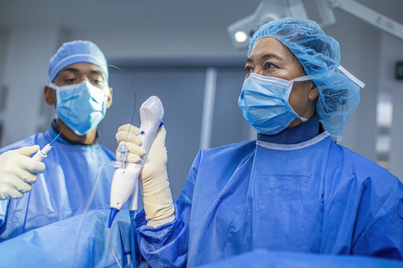
Ureteroscopy is one of the preferred methods to address small- to medium-sized stones located in the urinary tract.6 It does not require surgical cuts or incisions. Instead, once the patient is under anaesthesia, a urologist inserts a flexible, telescope-like instrument called a ureteroscope through the opening of the urinary tract and into the bladder. This scope functions as a camera to examine the urinary tract, locate the kidney stone, and treat it with various tools inserted through the ureteroscope’s shaft. Stones can be fragmented or dusted by lasers, or extracted by endoscopic graspers or baskets.7
Reducing complication risks from kidney stones
During ureteroscopy, surgeons need to maintain a balance between surgical visualisation, procedure time and patient safety. While fluid irrigation may be used to improve visibility, it can also increase pressure within the kidneys, a condition known as high intrarenal pressure (IRP).8
A panel of European experts9, agreed that any IRP exceeding normal physiological levels is to be considered high, and that high IRP during ureteroscopy is cause for concern due to its link to increased patient complications.10, 11 Those at highest risk include patients with recurrent urinary tract infections, patients with severe comorbidities, female patients, patients with tight ureters, patients with diabetes and patients with a narrow pelvic-ureteric junction 12, 13
Meaningful innovation to advance patient care
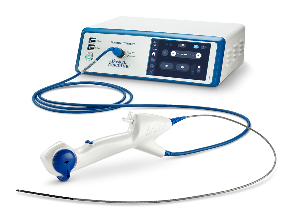
Until recently, urologists lacked a practical way to measure IRP in real time during ureteroscopy. The LithoVue™ Elite Single-Use Digital Flexible Ureteroscope System is one of the first ureteroscopes with a built-in pressure sensor. “This next-generation ureteroscope was developed in response to feedback from urologists and expert consensus on the importance of maintaining low intrarenal pressure for patient safety,” explains Miguel Aragon, vice president of Urology in EMEA at Boston Scientific. “It exemplifies our commitment to developing clinical solutions that enhance patient care while improving procedural efficiency and surgical decision-making.”
The LithoVue™ Elite Single-Use Digital Flexible Ureteroscope System recently obtained CE Mark. Boston Scientific will commence limited market release in the coming weeks.
Click here to learn more about LithoVue Elite

1 Data based on prevalence in absolute term in the following countries: Italy (Prezioso D, Illiano E, Piccinocchi G, Cricelli C, Piccinocchi R, Saita A, et al. Urolithiasis in Italy: An epidemiological study. Arch Ital di Urol e Androl. 2014;86: 99–102.doi:10.4081/aiua.2014.2.99); Spain (Morales-Martínez A, Melgarejo-Segura MT, Arrabal-Polo MA. Urinary stone epidemiology in Spain and worldwide. Arch Esp Urol. 2021;74: 1–3); France (Daudon M. Epidemiology of nephrolithiasis in France. Ann Urol (Paris). 2005;39: 209–231. doi:10.1016/j.anuro.2005.09.007); UK (Heers H, Turney BW. Trends in urological stone disease: a 5-year update of hospital episode statistics. BJU Int. 2016;118: 785–789. doi:10.1111/bju.13520); Germany (2. Fisang C, Anding R, Müller SC, Latz S, Laube N. Urolithiasis - An interdisciplinary diagnostic, therapeutic and secondary preventive challenge. Dtsch Arztebl Int. 2015;112: 83–91. doi:10.3238/arztebl.2015.0083)
2 Ratkalkar VN, Kleinman JG. Mechanisms of Stone Formation. Clinical reviews in bone and mineral metabolism.2011; 9(3-4), 187. https://doi.org/10.1007/s12018-011-9104-8
3 New F, Somani BK. A Complete World Literature Review of Quality of Life (QOL) in Patients with Kidney Stone Disease (KSD). Curr Urol Rep. 2016;17: 1–6. doi:10.1007/s11934-016-0647-6
4 Fisang C, Anding R, Müller SC, Latz S, Laube N. Urolithiasis - An interdisciplinary diagnostic, therapeutic and secondary preventive challenge. Dtsch Arztebl Int. 2015;112: 83–91. doi:10.3238/arztebl.2015.0083
5 Skolarikos A, Neisius A, Petřík A, Somani B, Thomas K, Gambaro G. EAU guidelines on urolithiasis. Eur Assoc Urol. 2022.
6 EAU Patient information. Ureteroscopy (fURS). https://patients.uroweb.org/treatments/ureteroscopy/
7 Johns Hopkins Medicine. Ureteroscopy. Available: https://www.hopkinsmedicine.org/health/treatment-tests-and-therapies/ureteroscopy
8 Somani, B., N. Davis, E. Emiliani, M. I. Gökce, H. U. Jung, E. X. Keller, A. Miernik et al. Expert consensus on high intra-renal pressure during ureteroscopy: A pan-European delphi panel. European Urology 85 (2024): S1294.
9 See 8
10 Proietti S, Dragos L, Somani B, et al. In vitro comparison of maximum pressure developed by irrigation systems in a kidney model. J Endourol. 2017 May;31(5):522-7;
11 Tokas T, Herrmann TRW, Skolarikos A, et al. Pressure matters: intrarenal pressures during normal and pathological conditions, and impact of increased values to renal physiology. World J Urol. 2019 Jan;37(1):125-31.
12 Chugh, S., Pietropaolo, A., Montanari, E., Sarica, K., & Somani, B. K. (2020). Predictors of urinary infections and urosepsis after ureteroscopy for stone disease: a systematic review from EAU section of urolithiasis (EULIS). Current urology reports, 21, 1-8.
13 Bhojani, N., Miller, L. E., Bhattacharyya, S., Cutone, B., & Chew, B. H. (2021). Risk factors for urosepsis after ureteroscopy for stone disease: a systematic review with meta-analysis. Journal of endourology, 35(7), 991-1000.
* After shock wave lithotripsy, doctor might decide to place a ureteral stent
*** Learn more about ureteroscopy